
Long upper-arm plaster cast until wound healing is achieved. In all cases, suture repair of the periosteum is advisable. In older children (& amp amp amp amp amp gt or = 5 years of age) or in cases requiring compression radial screw fixation is recommended. In smaller children (& amp amp amp amp amp lt 5 years of age) fixation with Kirschner wires. Open reduction of the lateral humeral condyle via a lateral approach to the elbow joint. Incomplete, so-called hanging fractures of the lateral humeral condyle without notable secondary dislocation on follow-up. Relative: complete fractures of the lateral humeral condyle which demonstrate a dislocation & amp amp amp amp amp lt or = 2 mm on follow-up. Absolute: fractures with a complete dislocation or those in which plaster-free control X-ray on day 4 shows a gap of & amp amp amp amp amp gt 2 mm. In general, the prognosis is good with the majority of fractures healing well with little functional loss.Surgical treatment of lateral humeral condyle fractures with reduction and retention in order to prevent lasting malalignment, pseudarthrosis, and joint instability. Hemi-arthroplasty is also an option especially for three and four-part fractures, where the risk of malunion and avascular necrosis are high 1. Having said this, in almost all cases undisplaced fractures are treated conservatively, whereas operative open reduction and internal fixation (with a variety of intramedullary nails, plates and screws and K-wires) is reserved for displaced fractures 1. For example, someone who lives alone may not be able to do so without the use of one arm.

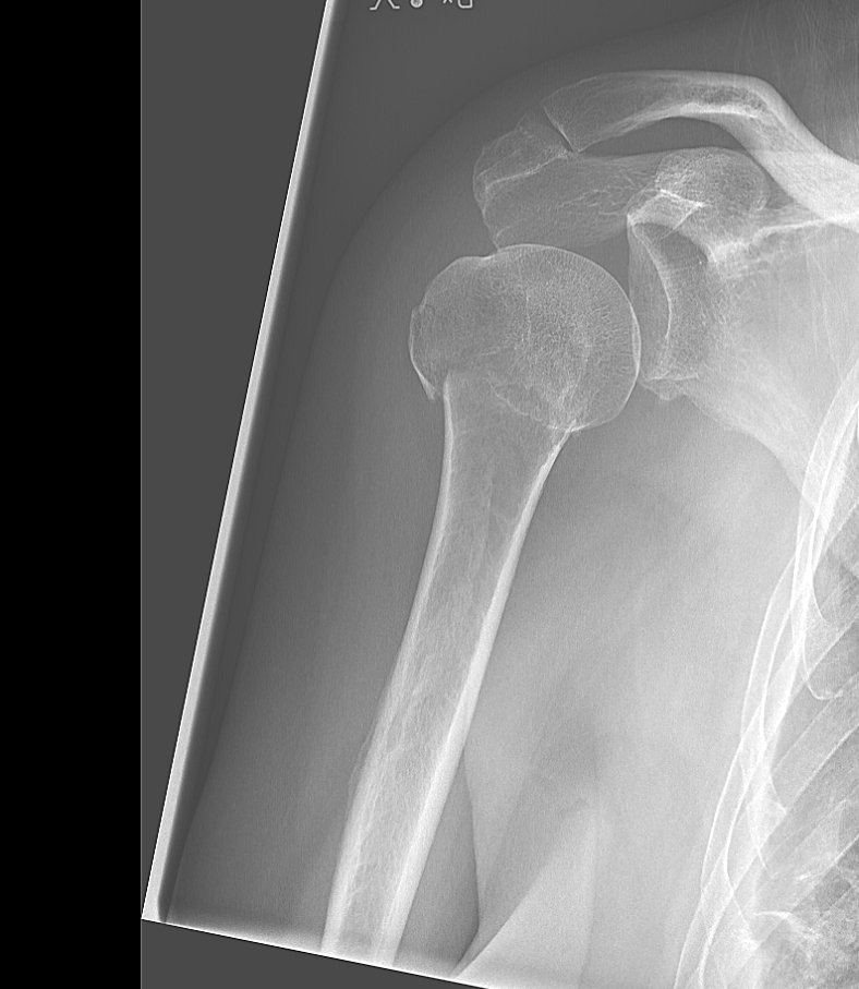
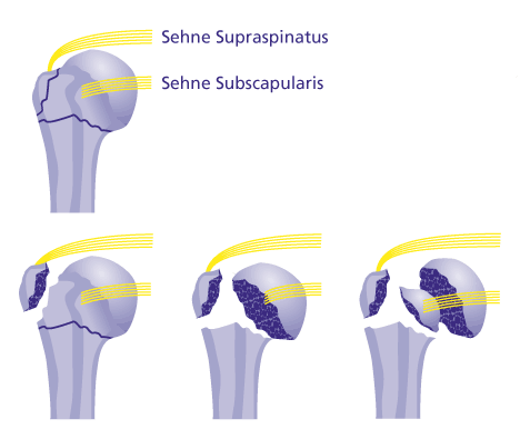
Management depends not only on the type of fracture but also importantly on the functional status and living situation of the patient. distal radial fracture (especially Colles fracture).presence of involvement of the articular surface.displacement and angulation of each part (>1 cm and >45 degrees respectively is particularly important in the Neer classification).fracture: it should be noted that in most instances a description of the fracture rather than a specific classification is sufficient, but the features required to classify the fracture should be included.In addition to reporting the presence of a fracture, it is important to assess and comment on a number of other features. The fracture is usually evident as a lucency and cortical breach with variable degrees of angulation, impaction and displacement. Regardless of the imaging performed, the number of displaced fragments should be assessed, to enable appropriate classification of the fracture ( Neer classification or AO classification are most commonly used). Additionally, CT (and especially 3D surface shaded reconstructions) has been shown to improve interobserver agreement on classification of proximal humeral fractures 4. CT can be useful if adequate views cannot be obtained, if fractures are unusual or if other fractures (e.g. Plain films are usually sufficient to characterize proximal humeral fractures, and thus to determine management. These forces may be compressive, tension, torsion or bending. Proximal humeral fractures usually result from a fall on an outstretched arm. Indirect forces transmitted through the proximal humerus and shoulder are the cause of most fractures. However, patients may present following a seizure, electrical shock or following direct trauma.

Younger patients usually present following a high-trauma incident, e.g. Many older patients present following a relatively innocuous fall. Most of these (90%) occur at home due to a fall, and in most cases they are an isolated injury 1. The majority of proximal humeral fractures occur in the elderly (mean age 65 years) with ~70% occurring in women, presumably due to the greater incidence of osteoporosis 1. As with other injuries, there is a bimodal distribution with a small peak amongst the young. They are most common in older populations and especially in those who are osteoporotic. Proximal humeral fractures represent around 5% of all fractures ?.


 0 kommentar(er)
